The sliding filament theory explains how a skeletal muscle contracts: thin actin filaments slide over thick myosin filaments, pulling the ends of each sarcomere closer together, while the filaments themselves do not change length. It is one of the most frequently asked topics from Locomotion and Movement in NEET, usually as a question on which band shortens, the role of calcium ions, or the order of events. This page explains the theory step by step, with two simple diagrams, quick revision points and practice questions at the end.
Sliding Filament Theory at a Glance
- What it explains: contraction of skeletal muscle fibres
- Main idea: thin (actin) filaments slide over thick (myosin) filaments towards the centre of the sarcomere
- Trigger: a nerve signal releases acetylcholine at the neuromuscular junction
- Key ion: Ca²⁺ released from the sarcoplasmic reticulum
- Energy source: ATP, hydrolysed by the myosin head
- What shortens: the sarcomere, the I band and the H zone
- What stays the same: the A band, and the length of both filaments
The Sarcomere: Where Contraction Happens
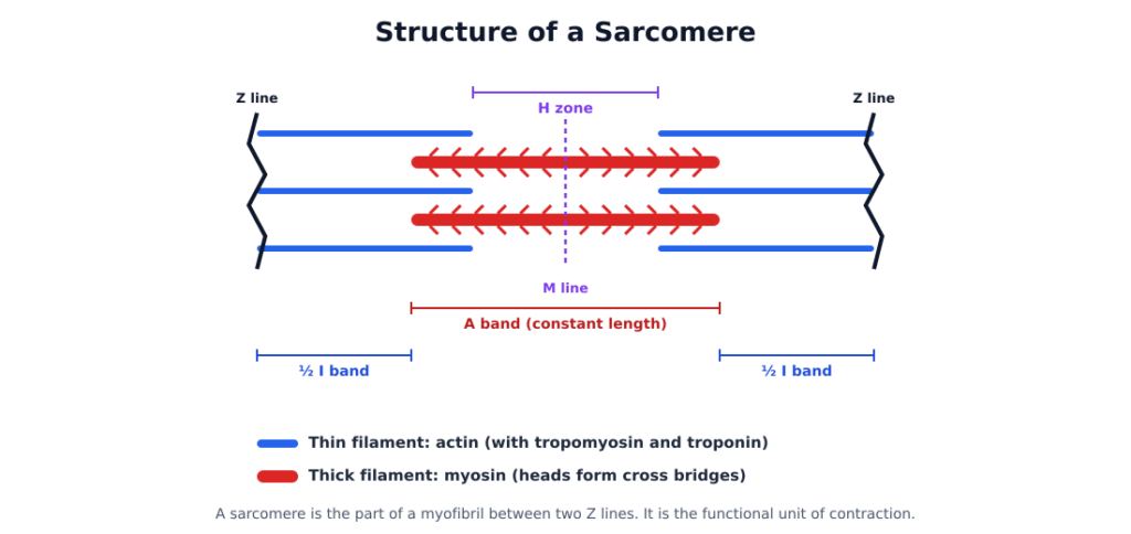
Each muscle fibre is packed with myofibrils, and each myofibril is a chain of repeating units called sarcomeres. A sarcomere is the part of a myofibril between two Z lines, and it is the functional unit of contraction.
| Part | What it is |
|---|---|
| Thin filament | Actin, with two regulatory proteins: tropomyosin and troponin |
| Thick filament | Myosin. Each myosin molecule has a globular head (heavy meromyosin) and a tail (light meromyosin) |
| Z line | An elastic line in the middle of each I band. The thin filaments are attached to it |
| I band (isotropic, light band) | Contains only thin filaments |
| A band (anisotropic, dark band) | The full length of the thick filaments, including the parts where thin filaments overlap them |
| H zone | The central part of the A band, where thin filaments do not overlap the thick filaments |
| M line | A thin line in the centre of the A band that holds the thick filaments together |
Two features of the proteins matter for contraction:
- In a resting muscle, troponin and tropomyosin cover the active sites on actin where myosin would bind.
- The myosin head is an ATPase enzyme. It has binding sites for ATP and for the active sites on actin.
Sliding Filament Theory Step by Step
1. The nerve signal reaches the muscle
A signal from the central nervous system travels down a motor neuron. At the neuromuscular junction (motor end plate), the neuron releases the neurotransmitter acetylcholine. This generates an action potential in the sarcolemma, which spreads through the muscle fibre.
2. Calcium ions are released
The action potential causes the sarcoplasmic reticulum to release Ca²⁺ ions into the sarcoplasm.
3. The active sites on actin are uncovered
Ca²⁺ binds to a subunit of troponin on the thin filament. This changes the position of troponin and tropomyosin, and the active sites on actin are uncovered.
4. Cross bridges form
Using energy from the hydrolysis of ATP, the myosin head binds to the exposed active site on actin, forming a cross bridge.
5. The power stroke
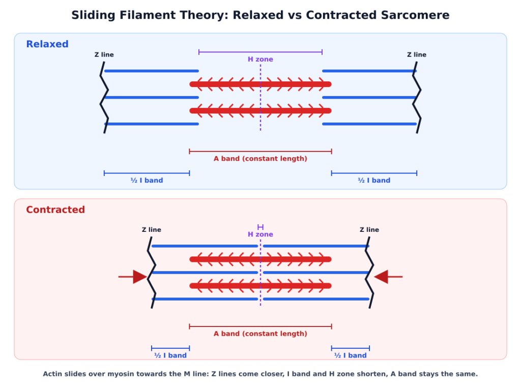
The cross bridge pulls the attached actin filament towards the centre of the A band. The Z lines attached to the actin are pulled inward too, so the sarcomere shortens. This is contraction.
6. The cross bridge breaks
The myosin head releases ADP and Pi and goes back to its relaxed state. A new ATP molecule binds to the head, and the cross bridge is broken.
7. The cycle repeats
The ATP is hydrolysed again, and the myosin head binds to the next active site further along the actin. Cross bridges keep forming and breaking, pulling the thin filaments further in, for as long as Ca²⁺ and ATP are available.
8. Relaxation
When the nerve signal stops, Ca²⁺ is pumped back into the sarcoplasmic reticulum. Troponin and tropomyosin cover the active sites on actin again, no new cross bridges can form, and the Z lines return to their original positions. The muscle relaxes.
What Changes During Contraction
| Part | During contraction |
|---|---|
| Sarcomere (Z line to Z line) | Shortens |
| I band | Shortens |
| H zone | Shortens, and may disappear |
| A band | Stays the same length |
| Length of actin and myosin filaments | Stays the same. They only slide past each other |
This is the most common NEET question on the topic. The A band keeps its length because it equals the length of the thick filament, which does not change.
Muscle Fatigue and Red and White Fibres
Repeated activation of a muscle can lead to the build-up of lactic acid, from the anaerobic breakdown of glycogen. This causes fatigue.
Muscles also differ in how they get their energy:
- Red fibres have a lot of myoglobin (a red, oxygen-storing pigment), plenty of mitochondria and a good blood supply. They are aerobic muscles.
- White fibres have very little myoglobin and fewer mitochondria, but a large sarcoplasmic reticulum. They depend on anaerobic respiration.
Quick Revision Points for NEET
- The sarcomere, between two Z lines, is the functional unit of contraction.
- Actin, with troponin and tropomyosin, makes the thin filament. Myosin makes the thick filament.
- Acetylcholine is the neurotransmitter at the neuromuscular junction.
- Ca²⁺ is released from the sarcoplasmic reticulum and binds to troponin.
- The myosin head is an ATPase, and it binds actin to form the cross bridge.
- Actin is pulled towards the centre of the A band (M line).
- The I band and H zone shorten. The A band does not.
- A new ATP is needed to break the cross bridge.
- Relaxation happens when Ca²⁺ is pumped back into the sarcoplasmic reticulum.
Practice Questions on the Sliding Filament Theory
1. During muscle contraction, which of the following does not change in length?
2. Ca²⁺ ions released during muscle contraction bind to
3. Ca²⁺ ions for muscle contraction are released from the
4. The myosin head acts as an enzyme. It is
5. In a contracting sarcomere, the thin filaments are pulled towards the
6. The neurotransmitter released at the neuromuscular junction to start muscle contraction is
Answers
- (C) A band. The A band equals the length of the thick filament, which does not change. The I band, H zone and sarcomere all shorten.
- (D) troponin. Ca²⁺ binds to a subunit of troponin, which moves troponin and tropomyosin and uncovers the active sites on actin.
- (B) sarcoplasmic reticulum. The sarcoplasmic reticulum of a muscle fibre stores Ca²⁺ and releases it when an action potential arrives.
- (B) an ATPase. The myosin head hydrolyses ATP and uses the energy to bind actin and pull it.
- (C) centre of the A band. Cross bridges pull the actin filaments towards the middle of the A band, which brings the Z lines closer.
- (B) acetylcholine. Acetylcholine released at the motor end plate generates the action potential in the sarcolemma.
Keep Practising
The sliding filament theory is one section of Locomotion and Movement. Revise the full chapter, including the types of movement, the skeletal system, joints and disorders of the muscular and skeletal system, from the Locomotion and Movement Class 11 notes. Then solve the Locomotion and Movement NEET PYQs to see how NEET asks this topic. The next chapter, Neural Control and Coordination, explains how the nerve signal that starts contraction is produced. For other chapters, see all Class 11 and 12 Biology notes.
Frequently Asked Questions
Q: What is the sliding filament theory in simple words? A: It says that a muscle contracts because the thin actin filaments slide over the thick myosin filaments towards the centre of each sarcomere. The filaments don’t shorten. They only overlap more, so the sarcomere becomes shorter.
Q: Which band does not change during muscle contraction? A: The A band. It equals the length of the thick (myosin) filament, which stays the same. The I band and the H zone become shorter.
Q: What is the role of calcium in muscle contraction? A: Ca²⁺ released from the sarcoplasmic reticulum binds to troponin. This moves troponin and tropomyosin off the active sites on actin, so myosin heads can bind and form cross bridges.
Q: Why is ATP needed in muscle contraction? A: The myosin head hydrolyses ATP to get the energy to bind actin and pull it. A new ATP is also needed to break the cross bridge, so that the head can bind again further along.
Q: Who proposed the sliding filament theory? A: It was proposed in 1954 by two research teams working independently: Andrew Huxley and Rolf Niedergerke, and Hugh Huxley and Jean Hanson.