
Quick answer: A locomotion and movement NEET PYQ analysis shows that NTA asks about two questions a year from this chapter, and they come from four areas: the sliding filament theory of muscle contraction (bands, Ca²⁺, troponin, myosin heads), the human skeletal system (vertebrae, ribs, girdles and bone counts), types of joints with examples, and disorders of the muscular and skeletal system (myasthenia gravis, tetany, gout, osteoporosis). The 2026 papers asked six questions from these areas.
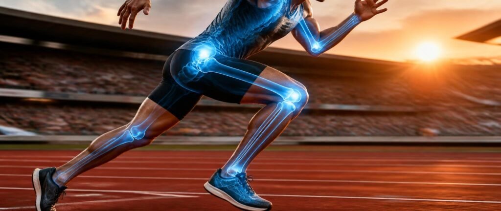
This guide breaks down what NTA repeats, the tables to memorise and PYQ-pattern questions with answers. For chapter notes, use our Locomotion and Movement notes.
Want to test yourself first? Solve the Locomotion and Movement PYQs for free, then use this guide to fix the gaps.
Table of Contents
Why This Chapter Is Reliable Marks
Locomotion questions are almost all counts and matches: which band stays the same, how many true ribs, which joint is between the atlas and axis, which disorder comes from uric acid. Learn those tables and the chapter becomes easy marks. To see where it sits among other chapters, check the NEET 2027 chapter weightage.
Muscle tissue types are covered in our Structural Organisation PYQ analysis, and the nerve impulse that starts a contraction is in our Neural and Endocrine PYQ analysis. The sarcomere is also a common diagram question, so our guide to diagram-based biology questions is worth a look.
Locomotion and Movement NEET PYQ Analysis: What NTA Repeats
When you sort the past NEET biology PYQs from this chapter, four question types cover nearly everything.
| Question type | Frequency | What it tests |
|---|---|---|
| Contraction | Most frequent | Sarcomere bands, actin and myosin, Ca²⁺ and troponin, steps of the sliding filament theory |
| Skeleton | Very frequent | Bone counts, vertebrae, ribs, girdles, limb bones |
| Joints | Very frequent | Fibrous, cartilaginous and synovial joints with examples |
| Disorders | Regular | Myasthenia gravis, muscular dystrophy, tetany, arthritis, osteoporosis, gout |
Types of Movement
| Type | Example |
|---|---|
| Amoeboid | Macrophages and leucocytes in blood; uses pseudopodia formed by cytoplasmic streaming and microfilaments |
| Ciliary | Cilia in the trachea remove dust; movement of the ovum through the female reproductive tract |
| Muscular | Movement of limbs, jaws and tongue |
Muscle Structure
Muscles are of mesodermal origin and make up about 40–50% of the body weight of an adult human.
| Term | Meaning |
|---|---|
| Fascicle | Muscle bundle, held together by connective tissue called fascia |
| Sarcolemma | Plasma membrane of a muscle fibre |
| Sarcoplasm | Cytoplasm of a muscle fibre |
| Sarcoplasmic reticulum | Endoplasmic reticulum that stores Ca²⁺ |
| Sarcomere | The part of a myofibril between two successive Z lines; the functional unit of contraction |
Bands of a sarcomere:
| Band or line | What it contains |
|---|---|
| A band (dark, anisotropic) | Thick myosin filaments |
| I band (light, isotropic) | Thin actin filaments |
| Z line | Bisects the I band; actin filaments are attached to it |
| M line | Thin membrane in the middle of the A band holding the thick filaments |
| H zone | Central part of the A band not overlapped by thin filaments |
Contractile Proteins
- Actin (thin filament): two helically wound F-actin strands, each a polymer of G-actin monomers. Tropomyosin runs along the length, and troponin sits at regular intervals; in a resting muscle, troponin masks the binding sites for myosin on actin.
- Myosin (thick filament): a polymer of meromyosin. Each meromyosin has a globular head with a short arm (heavy meromyosin) and a tail (light meromyosin). The head, called the cross arm, is an active ATPase with binding sites for ATP and actin.
The Sliding Filament Theory
The sliding filament theory says that muscle contracts because thin filaments slide over thick ones.
- A signal from the CNS reaches the neuromuscular junction through a motor neuron. A motor neuron and the muscle fibres it supplies form a motor unit.
- Acetylcholine is released and starts an action potential in the sarcolemma.
- Ca²⁺ is released from the sarcoplasmic reticulum into the sarcoplasm.
- Ca²⁺ binds to troponin, unmasking the active sites on actin.
- Myosin heads, using energy from ATP hydrolysis, bind actin to form cross bridges and pull the actin filaments towards the centre of the A band.
- The Z lines are pulled inward, so the sarcomere shortens.
- ADP and Pi are released; a new ATP binds and the cross bridge breaks. The cycle repeats until Ca²⁺ is pumped back and the actin sites are masked again.
| During contraction | Change |
|---|---|
| A band | Stays the same length |
| I band | Shortens |
| H zone | Shortens or disappears |
| Sarcomere | Shortens |
Repeated activation leads to fatigue, as lactic acid builds up from the anaerobic breakdown of glycogen.
| Feature | Red fibres | White fibres |
|---|---|---|
| Myoglobin | High | Low |
| Mitochondria | Many | Few |
| Sarcoplasmic reticulum | Less | More |
| Energy | Aerobic | Anaerobic |
The Skeletal System: Numbers to Learn
The skeletal system of an adult human has 206 bones: 80 in the axial skeleton and 126 in the appendicular skeleton.
| Part | Facts |
|---|---|
| Skull | 22 bones (8 cranial, 14 facial); with the U-shaped hyoid and 6 ear ossicles, 29 bones in the skull region; dicondylic (two occipital condyles) |
| Vertebral column | 26 vertebrae: cervical 7, thoracic 12, lumbar 5, sacral 1 (fused), coccygeal 1 (fused); the first vertebra, the atlas, articulates with the occipital condyles |
| Ribs | 12 pairs; each rib is bicephalic (two articulation surfaces at its dorsal end) |
| True ribs | First 7 pairs, attached to the vertebral column and to the sternum by hyaline cartilage |
| False (vertebrochondral) ribs | 8th, 9th and 10th pairs, joined to the 7th rib’s cartilage instead of the sternum |
| Floating ribs | 11th and 12th pairs, not joined ventrally |
Almost all mammals, including humans, have seven cervical vertebrae.
Appendicular skeleton:
| Part | Bones |
|---|---|
| Forelimb (30 bones) | Humerus, radius and ulna, 8 carpals, 5 metacarpals, 14 phalanges |
| Hindlimb (30 bones) | Femur (longest bone), tibia and fibula, 7 tarsals, 5 metatarsals, 14 phalanges, patella (knee cap) |
| Pectoral girdle | Clavicle (collar bone) and scapula; the scapula’s glenoid cavity forms the shoulder joint with the humerus |
| Pelvic girdle | Two coxal bones, each formed by fusion of the ilium, ischium and pubis; the femur fits into the acetabulum; the halves meet at the pubic symphysis |
Joints
| Joint type | Movement | Example |
|---|---|---|
| Fibrous | None | Sutures between flat skull bones |
| Cartilaginous | Limited | Between adjacent vertebrae |
| Synovial: ball and socket | Wide | Between the humerus and the pectoral girdle (shoulder) |
| Synovial: hinge | One plane | Knee joint |
| Synovial: pivot | Rotation | Between the atlas and axis |
| Synovial: gliding | Sliding | Between the carpals |
| Synovial: saddle | Two planes | Between the carpal and metacarpal of the thumb |
Joints questions were asked in 2023 and twice in 2024, mostly as matching questions.
Disorders of the Muscular and Skeletal System
| Disorder | Cause or feature |
|---|---|
| Myasthenia gravis | Autoimmune disorder of the neuromuscular junction; fatigue, weakening and paralysis of skeletal muscle |
| Muscular dystrophy | Progressive degeneration of skeletal muscle, mostly genetic |
| Tetany | Rapid spasms (wild contractions) due to low Ca²⁺ in body fluid |
| Arthritis | Inflammation of joints |
| Osteoporosis | Age-related loss of bone mass and higher chance of fractures; low oestrogen is a common cause |
| Gout | Inflammation of joints due to accumulation of uric acid crystals |
Worked PYQ-Pattern Questions
Q1. During muscle contraction, which band does not change in length? (a) I band (b) H zone (c) A band (d) Sarcomere
Answer: (c) A band. The I band and H zone shorten.
Q2. In a resting muscle, the active sites for myosin on actin are masked by:
Answer: Troponin. Ca²⁺ binds to troponin to unmask them.
Q3. How many pairs of true ribs are there in humans?
Answer: Seven pairs.
Q4. The joint between the atlas and the axis is a: (a) Hinge joint (b) Pivot joint (c) Gliding joint (d) Saddle joint
Answer: (b) Pivot joint.
Q5. Gout is caused by:
Answer: Accumulation of uric acid crystals in the joints, causing inflammation.
Q6. Which bones fuse to form each coxal bone of the pelvic girdle?
Answer: Ilium, ischium and pubis.
Common Traps in This Chapter
- Saying the A band shortens during contraction
- Saying Ca²⁺ binds to tropomyosin or myosin (it binds to troponin)
- Counting the 8th–10th ribs as true ribs
- Calling the knee a ball and socket joint
- Mixing up tetany (low Ca²⁺) and myasthenia gravis (autoimmune)
- Writing 33 vertebrae for adults (the adult count is 26, after fusion)
Practise Every Locomotion and Movement PYQ
This locomotion and movement NEET PYQ analysis shows you where the marks are. Solving real past questions makes the counts and matches automatic. Every Locomotion and Movement previous year question is available chapter-wise, free:
👉 Solve all Locomotion and Movement NEET PYQs
Keep the sarcomere, skeleton and joints tables beside you for the first round, then solve the second round without them.
Conclusion
Locomotion and Movement is a chapter of numbers and matches. Use this locomotion and movement NEET PYQ analysis to master the sliding filament theory, the bone counts, joints and disorders. Then practise the past questions until each answer comes without thinking.
FAQ
Q: How many questions come from Locomotion and Movement in NEET? A: Usually about two questions a year, on muscle contraction, the skeleton, joints or disorders.
Q: What is the most important topic in this chapter for NEET? A: Muscle contraction is the most tested, especially the sarcomere bands and the role of Ca²⁺ and troponin. The skeletal system comes next.
Q: Which band stays constant during muscle contraction? A: The A band. The I band and H zone shorten as the actin filaments slide inward.
Q: How many bones are there in the human body? A: 206 bones in an adult: 80 in the axial skeleton and 126 in the appendicular skeleton.
Q: What causes tetany? A: Low Ca²⁺ levels in body fluid, which cause rapid spasms in muscles.
Q: Where can I practise Locomotion and Movement NEET PYQs for free? A: You can solve every Locomotion and Movement previous year question chapter-wise for free using the practice links in this article.
Related posts:
 Animal Kingdom NEET PYQ Analysis: Examples NTA Asks Again and Again
 Plant Kingdom NEET PYQ Analysis: Algae, Bryophytes and Gymnosperms Decoded
 Body Fluids and Circulation NEET PYQ Analysis: ECG, Cardiac Cycle and Blood
 Breathing and Exchange of Gases NEET PYQ Analysis: Volumes, Transport and Regulation