
Quick answer: A structural organisation in animals NEET PYQ analysis shows that NTA asks about two questions a year from this chapter, and they come from three areas: animal tissues (types and locations of epithelium, connective tissue, muscle and cell junctions), cockroach anatomy (mouthparts, digestive and excretory organs, reproductive system and sexual differences) and frog anatomy (heart, respiration, excretion, brain and reproduction). Most questions match a tissue or organ with its location or function.
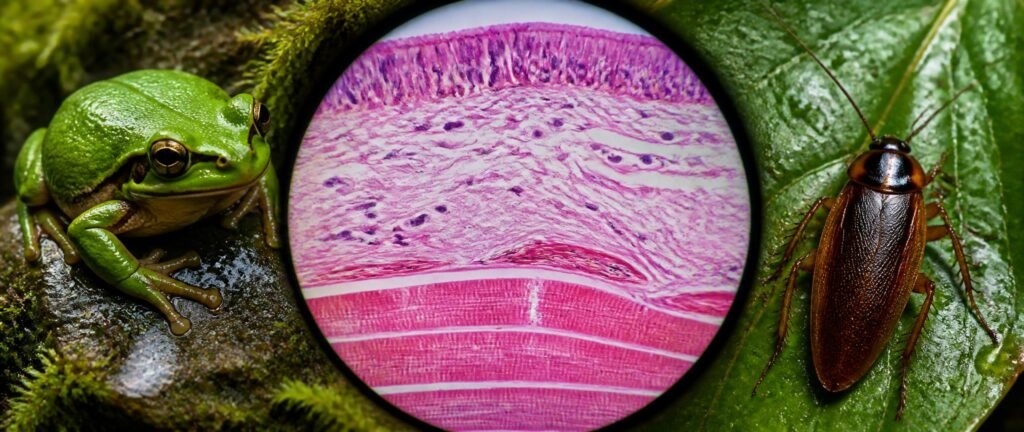
This guide breaks down what NTA repeats, the tables you must know and PYQ-pattern questions with answers. For chapter notes, use our Structural Organisation in Animals notes.
Want to test yourself first? Solve the Structural Organisation in Animals PYQs for free, then use this guide to fix the gaps.
Table of Contents
Why This Chapter Is Easy Marks
The chapter is descriptive, and its questions keep returning to the same NCERT facts: where ciliated epithelium is found, what tendons connect, how many chambers the frog’s heart has, and which segment carries the cockroach’s anal styles. To see where it sits among other chapters, check the NEET 2027 chapter weightage.
The current NEET syllabus asks for animal tissues and a brief account of the digestive, circulatory, respiratory, nervous and reproductive systems of a representative animal. Past papers have asked about both the cockroach and the frog, so this guide covers both. Our Animal Kingdom PYQ analysis covers the classification side, and our Body Fluids PYQ analysis builds on the blood and heart facts here.
Structural Organisation in Animals NEET PYQ Analysis: What NTA Repeats
When you sort the past NEET biology PYQs from this chapter, three question types cover nearly everything.
| Question type | Frequency | What it tests |
|---|---|---|
| Animal tissues | Most frequent | Epithelium types and locations, connective tissue, muscle types, glands, cell junctions |
| Cockroach anatomy | Very frequent | Mouthparts, wings, gizzard, hepatic caecae, Malpighian tubules, reproductive organs, anal cerci and styles |
| Frog anatomy | Very frequent | Three-chambered heart, cutaneous respiration, ureotelism, brain, sexual dimorphism |
For a quick warm-up, try these five quick Structural Organisation questions.
Epithelial Tissue
| Type | Structure | Found in |
|---|---|---|
| Squamous | Single thin layer of flat cells | Walls of blood vessels, air sacs of lungs |
| Cuboidal | Single layer of cube-like cells | Ducts of glands, tubular parts of nephrons (microvilli in the PCT) |
| Columnar | Tall, slender cells; may have microvilli | Lining of the stomach and intestine |
| Ciliated | Columnar or cuboidal cells with cilia | Bronchioles and fallopian tubes, to move particles or mucus |
| Compound (stratified) | Several layers | Dry skin surface, moist buccal cavity and pharynx, inner lining of salivary and pancreatic ducts |
Glands: goblet cells of the alimentary canal are unicellular glands; salivary glands are multicellular. Exocrine glands release mucus, saliva, earwax, oil, milk and digestive enzymes through ducts. Endocrine glands have no ducts and release hormones into the fluid around them.
Cell junctions:
| Junction | Role |
|---|---|
| Tight junctions | Stop substances leaking across a tissue |
| Adhering junctions | Cement neighbouring cells together |
| Gap junctions | Connect the cytoplasm of adjacent cells for rapid transfer of ions and small molecules |
Connective Tissue
| Type | Key facts |
|---|---|
| Areolar (loose) | Beneath the skin; contains fibroblasts, macrophages and mast cells |
| Adipose (loose) | Below the skin; stores fat |
| Dense regular | Tendons attach muscle to bone; ligaments attach bone to bone |
| Dense irregular | Found in the skin |
| Cartilage | Cells called chondrocytes; in the tip of the nose, outer ear, between adjacent vertebrae, and in limbs and hands in adults |
| Bone | Hard matrix of calcium salts and collagen; bone cells (osteocytes) sit in lacunae |
| Blood | Fluid connective tissue |
Animal tissues questions often ask which cells make the fibres. Fibroblasts secrete collagen and elastin fibres, and every connective tissue except blood secretes fibres of structural proteins.
Muscle and Neural Tissue
| Muscle | Appearance | Control | Location |
|---|---|---|---|
| Skeletal | Striated | Voluntary | Attached to bones, such as the biceps |
| Smooth | Fusiform, no striations | Involuntary | Walls of blood vessels, stomach and intestine |
| Cardiac | Striated, branched | Involuntary | Heart; cell junctions fuse the plasma membranes so cells contract as a unit |
In neural tissue, neurons are the excitable cells, and neuroglia make up more than half the volume of neural tissue in the body.
Cockroach Anatomy: The Facts NTA Repeats
Periplaneta americana is brown or black, 34–53 mm long, with a head, thorax and abdomen.
| System or part | Key facts |
|---|---|
| Head and mouthparts | Triangular head; biting and chewing mouthparts: labrum (upper lip), mandibles, maxillae, labium (lower lip) and hypopharynx (tongue) |
| Wings | Forewings (tegmina) are mesothoracic, opaque, dark and leathery; hindwings are metathoracic, transparent and used in flight |
| Digestive system | Crop stores food; gizzard (proventriculus) has six chitinous teeth for grinding; a ring of 6–8 hepatic (gastric) caecae at the junction of foregut and midgut secretes digestive juice |
| Excretion | 100–150 Malpighian tubules at the junction of midgut and hindgut remove nitrogenous wastes; fat body, nephrocytes and urecose glands also help; the cockroach is uricotelic |
| Circulation | Open system; colourless haemolymph; tubular heart with 13 chambers and ostia |
| Respiration | Network of tracheae opening through 10 pairs of spiracles |
| Nervous system and eyes | Ganglia joined by nerve cords (3 in the thorax, 6 in the abdomen); compound eyes with about 2000 hexagonal ommatidia give mosaic vision |
Cockroach anatomy for reproduction:
| Feature | Male | Female |
|---|---|---|
| Gonads | A pair of testes in the 4th–6th abdominal segments | A pair of ovaries, each with eight ovarioles |
| Accessory organs | Mushroom-shaped gland in the 6th–7th segments; sperm stored in spermatophores | Spermatheca in the 6th segment |
| Anal cerci | Present (10th segment) | Present |
| Anal styles | Present (9th sternum) | Absent |
Fertilised eggs are packed in an ootheca, a dark reddish to blackish-brown capsule about 8 mm long. A female produces 9–10 oothecae, each with 14–16 eggs. Development is paurometabolous, through nymph stages that moult about 13 times.
Frog Anatomy: The Facts NTA Repeats
| System or part | Key facts |
|---|---|
| Habits | Cold-blooded; shows camouflage; avoids extreme heat and cold by aestivation (summer sleep) and hibernation (winter sleep) |
| Skin | Moist and slimy; the frog never drinks water but absorbs it through the skin |
| Sexual dimorphism | Males have vocal sacs and a copulatory pad on the first digit of the forelimbs |
| Digestive system | Short alimentary canal (carnivore); bilobed tongue; bile from the liver is stored in the gall bladder |
| Respiration | Through the skin in water (cutaneous); through the buccal cavity, skin and lungs on land; only through the skin during aestivation and hibernation |
| Circulation | Closed; three-chambered heart (two atria, one ventricle) with a sinus venosus and conus arteriosus; hepatic portal and renal portal systems; nucleated RBCs |
| Excretion | Ureotelic; in males, ureters act as urinogenital ducts; in females, ureters and oviducts open separately into the cloaca |
| Brain | Forebrain (olfactory lobes, cerebral hemispheres, diencephalon), midbrain (a pair of optic lobes), hindbrain (cerebellum, medulla oblongata); 10 pairs of cranial nerves |
| Sense organs | Tympanum receives sound; a nictitating membrane protects the eye in water |
| Reproduction | Female lays 2500–3000 ova at a time; fertilisation is external, in water; development through a tadpole larva (metamorphosis) |
Frog anatomy questions most often ask about the three-chambered heart, cutaneous respiration and the ureotelic nature of the frog.
Worked PYQ-Pattern Questions
Q1. Ciliated epithelium is found in: (a) Walls of blood vessels (b) Bronchioles and fallopian tubes (c) Ducts of glands (d) Skin surface
Answer: (b). The cilia move particles or mucus in one direction.
Q2. Tendons connect:
Answer: Muscle to bone. Ligaments connect bone to bone.
Q3. In the cockroach, anal styles are found in: (a) Both sexes (b) Males only (c) Females only (d) Nymphs only
Answer: (b) Males only. Anal cerci are present in both sexes.
Q4. Malpighian tubules in the cockroach are found at the junction of:
Answer: The midgut and hindgut. They remove nitrogenous wastes.
Q5. The heart of a frog has:
Answer: Three chambers: two atria and one ventricle.
Q6. Which junction allows rapid transfer of ions between neighbouring cells? (a) Tight junction (b) Adhering junction (c) Gap junction (d) Desmosome
Answer: (c) Gap junction.
Common Traps in This Chapter
- Mixing up tendons (muscle to bone) and ligaments (bone to bone)
- Saying anal styles are present in female cockroaches
- Placing hepatic caecae at the midgut–hindgut junction (that is where the Malpighian tubules are)
- Calling the frog uricotelic (it is ureotelic; the cockroach is uricotelic)
- Saying the frog’s heart has four chambers
- Calling cardiac muscle voluntary or non-striated
Practise Every Structural Organisation in Animals PYQ
This structural organisation in animals NEET PYQ analysis shows you where the marks are. Solving real past questions makes the tissue and organ facts automatic. Every previous year question from this chapter is available chapter-wise, free:
👉 Solve all Structural Organisation in Animals NEET PYQs
Keep the tissue, cockroach and frog tables beside you for the first round, then solve the second round without them.
Conclusion
Structural Organisation in Animals is descriptive and highly predictable. Use this structural organisation in animals NEET PYQ analysis to master the tissue tables, cockroach anatomy and frog anatomy. Then practise the past questions until each location and function comes without thinking.
❓ FAQ
Q: How many questions come from Structural Organisation in Animals in NEET? A: Usually about two questions a year, from animal tissues, the cockroach or the frog.
Q: What is the most important topic in this chapter for NEET? A: Animal tissues are the most tested, especially the types and locations of epithelium, connective tissue and muscle. Cockroach anatomy comes next.
Q: Is the frog or the cockroach in the NEET syllabus? A: The syllabus asks for a brief account of the systems of a representative animal, and past papers have asked about both. Study both, focusing on the facts in this guide.
Q: What is the function of Malpighian tubules in the cockroach? A: They remove nitrogenous wastes from the haemolymph. The cockroach excretes uric acid, so it is uricotelic.
Q: What is the difference between a tendon and a ligament? A: A tendon attaches a skeletal muscle to a bone, while a ligament attaches one bone to another. Both are dense regular connective tissue.
Q: Where can I practise Structural Organisation in Animals NEET PYQs for free? A: You can solve every previous year question from this chapter chapter-wise for free using the practice links in this article.